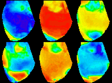

Regions of pacemaker cells in the heart
generate action potentials periodically due to a slow rise in their
membrane potential. The large regions of non-pacemaker cells in the
heart do not activate spontaneously, but action potentials are generated
when the front of a propagating wave passes through these cells. Action
potentials in the heart are characterized by a rapid (1-2 millisecond)
depolarization phase followed by a much slower (200-400 millisecond)
repolarization phase as the membrane potential recovers to resting
levels. These long action potentials are required to sustain the contraction
of the heart and force blood throughout the body.
During some abnormal conditions, such as lack of oxygen, very rapid
self-sustaining electrical activity can arise from abnormal pacemaker
cells or from the circulation of rotating waves of electrical activity
(often called reentrant waves, rotors, or spiral waves). Rapid activation
(tachycardia) prevents normal coordinated contractions of the heart
and can quickly (within 5 minutes) lead to death. The most dangerous
cardiac arrhythmia is ventricular fibrillation. Most tachycardias,
including fibrillation, result from spiral waves. Spiral waves have
been studied using numerical, theoretical, and experimental methods.
In the heart, the study of spiral waves requires high temporal and
spatial resolution, in addition to the ability to record from a large
number of sites. Video imaging technology, incorporating CCD, CMOS,
and photodiode array (PDA) cameras, is the most appropriate tool to
study spiral waves in the heart.
The articles contained in this library with links to the left touch
on some important issues to be considered when choosing a camera system
and planning an experiment. You will find information on the selection
of dyes, sources of noise, ways to
minimize noise and a link to our Ask
the Expert section. Although probably not complete, we consider
it valuable.
|
|


